The Midline of a Living Human Brain
 A
A
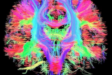
fountain of long cell fibers explodes along the midline of the human brain.
Computational neuroscientists study nervous systems in terms of their information processing capabilities. Standing at the junction of computer science and neuroscience, they have both the tools and the impetus to understand the details of the connectomes of whichever organisms they study.
An approach that has been taken in humans involves using the technique of diffusion tensor imaging, and MRI technique that can determine the direction that axons run in in an intact brain.
For example, the above image (by Thomas Schultz) shows a DTI-derived image of the connections that run through the midline of a living human brain.
Such images are of great potential use in studying brain lesions, doing studies on brain function, clinical diagnosis, and whole-brain level analysis of neural circuits. However, they lack the resolution needed to map individual synapses, thus falling short (for the time being) of being able to comprehensively map the connections between neurons in a brain. For this, we have to go to microscopy techniques that involve looking directly at neural tissue.
These can only been done in animals, because it is presently illegal to harvest brain tissue from humans for experimental purposes (again, a no-brainer.)
(Image by Thomas Schultz shows a DTI-derived image of the connections that run through the midline of a living human brain.)


